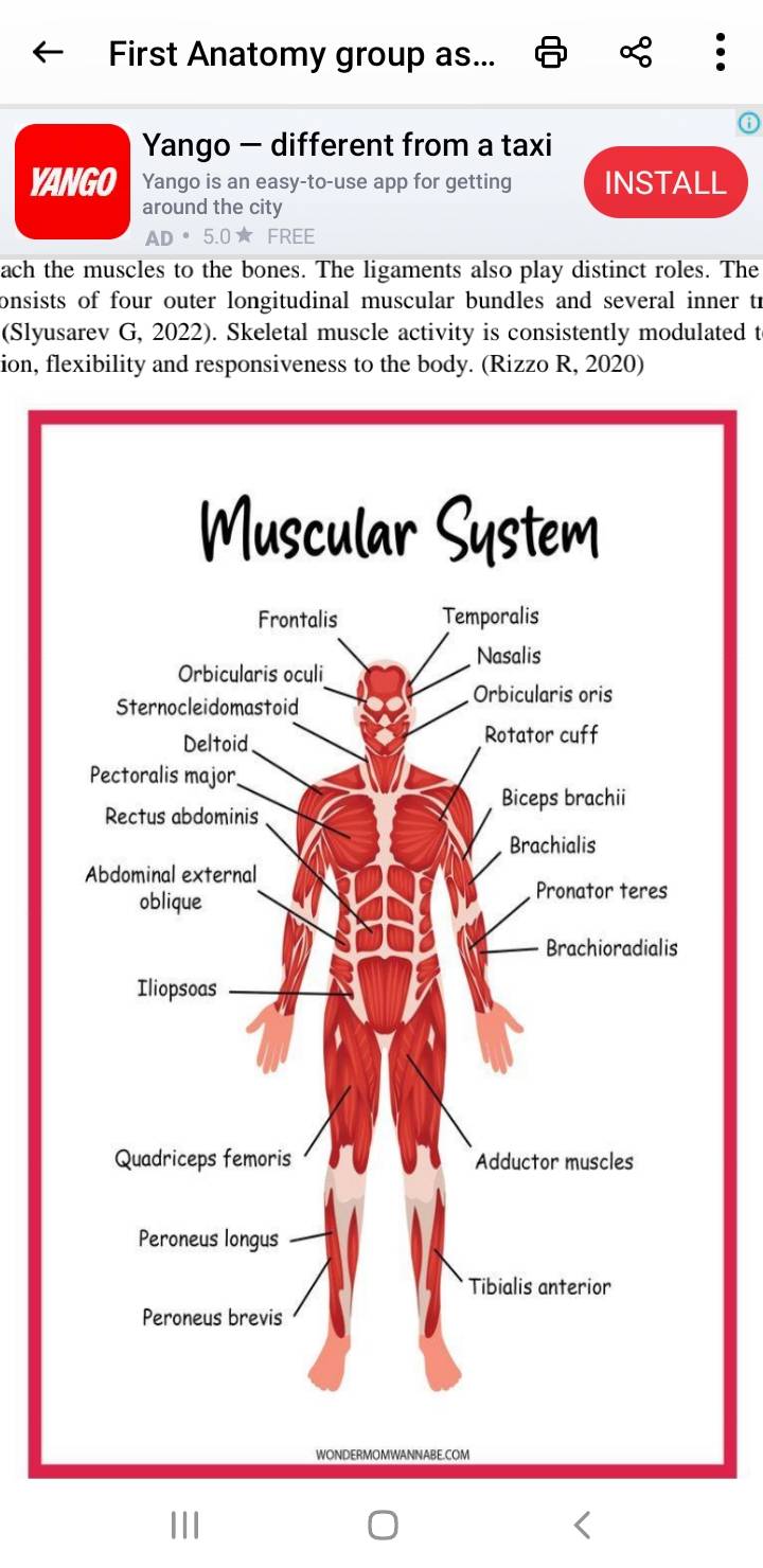
The muscular system is an organ system which is made up of specialized contractile tissues known as the muscle tissue. There are three types of muscle tissues which include: cardiac muscle which forms the muscular layer that surrounds the heart; smooth muscles which surrounds blood vessels and hollow organs and the skeletal muscle that attaches to the bones and provides voluntary motions.Apart from muscles, it contains the tendons which attach the muscles to the bones. The ligaments also play distinct roles. The muscular system consists of four outer longitudinal muscular bundles and several inner transversal muscles. . Skeletal muscle activity is consistently modulated to provide coordination, flexibility and responsiveness to the body.
GENERAL FUNCTIONS OF THE MUSCULAR SYSTEM.
MOBILITY: The basic role of the muscular system is to enable various types of movements. Major movements involve coordinated actions on a larger scale, such as walking, running and swimming. Fine movements also encompasses writing, speaking, facial expression among others.
STABILITY: Tendons in the knee joint and shoulder joint are so important for stabilization. Core muscles stabilize the body and help in activities such as weightlifting.
POSTURE: Skeletal muscles play a crucial role in maintaining proper body posture when sitting or standing.
CIRCULATION: The heart is a muscle that pumps blood to the various part of the body .The movement of the heart is involuntary and its contraction is stimulated by electrical signals. Smooth muscle in the arteries and veins plays a further role in the circulation of blood around the body.
RESPIRATION: The process of breathing relies on the diaphragm muscle.The diaphragm is a muscle shaped like a dome, positioned beneath the lungs. When the diaphragm contracts, it moves downwards, expanding the chest cavity. This action allows the lungs to fill up with air. On the other hand, when the diaphragm muscle relaxes, it assists in expelling air from the lungs. When individuals aim to take deeper breaths, additional muscles come into play. These include the muscles in the abdomen, back, and neck, providing assistance.
DIGESTION: Smooth muscles in the gastrointestinal or GI tract control digestion. The GI tract stretches from the mouth to the anus. Food moves through the digestive system with a wave-like motion called peristalsis. Muscles in the walls of the hollow organs contract and relax to cause this movement, which pushes food through the esophagus into the stomach.
URINATION: The urinary system consists of various types of muscles, including smooth and skeletal muscles, found in different parts such as the bladder, kidneys, penis or vagina, prostate, ureters, and urethra. These muscles, along with the nerves, collaborate to control the storage and release of urine from the bladder. Conditions related to the urinary system, such as difficulties in controlling the bladder or the inability to empty it completely, occur due to nerve damage that affects the communication between the nerves and muscles responsible for urinary functions.
CHILDBIRTH: During the process of childbirth, the smooth muscles within the uterus undergo expansion and contraction. These movements facilitate the movement of the baby through the vagina. Additionally, the pelvic floor muscles play a role in guiding the baby's head as it descends through the birth canal.
VISION: The eye is controlled by six skeletal muscles that are responsible for its movements. These muscles exhibit both speed and precision, enabling the eye to perform several functions:
Stabilizing the image: The muscles help in maintaining a steady image by adjusting the position of the eye.
Scanning the surroundings: The muscles allow the eye to scan and observe the surrounding area.
Tracking moving objects: These muscles enable the eye to track and follow objects in
motion.
When an individual sustains damage to their eye muscles, it can have a negative impact on their vision, potentially leading to visual impairments.
ORGAN PROTECTION: The muscles located in the torso serve the crucial function of safeguarding the internal organs positioned at the front, sides, and back of the body. In addition to the muscles, the spine bones and ribs offer an additional layer of protection.
Furthermore, these muscles play a role in shielding both the bones and organs by absorbing shocks and minimizing friction within the joints. This mechanism helps in reducing the impact and strain on the skeletal structure while promoting smooth movement and protecting the vital organs from potential damage.
TEMPERATURE REGULATION: The muscular system plays a vital role in regulating
the body's temperature. Approximately 85 percent of the heat generated within the body
is a result of muscle contractions. When the body's temperature drops below the optimal
level, the skeletal muscles increase their activity to generate heat. Shivering is an
example of this mechanism, where the muscles contract rapidly. Additionally, the
muscles in the blood vessels constrict to help maintain body heat. To restore the body
temperature to its normal range, the smooth muscles in the blood vessels relax. This
relaxation promotes increased blood flow, allowing excess heat to be released through
the skin. This process aids in regulating body temperature and maintaining a stable
internal environment.
FUNCTIONS OF THE INDIVIDUAL ORGANS OF THE MUSCULAR SYTSTEM.
The muscular system consist mainly of skeletal muscles, associated with tendons and other muscle – related structures. Here are the individual organs and their specific functions.
Skeletal muscles: Skeletal muscles are voluntary muscles attached to bones and tendons. Their main function is to generate force and produce movement, which allows us to perform various voluntary actions like walking, running and lifting. Examples are;
biceps brachii, quadriceps femoris, gastrocnemius, pectoralis major, gluteus maximus.
Smooth muscles: These are found in the walls of internal organs, such as the digestive tract, blood vessels and respiratory tract. They are involuntary muscles and are responsible for movements within these organs, like peristalsis (wave- like contractions) in the digestive system or dilation of blood vessels. Examples are; smooth muscles in the walls of the digestive tract, blood vessels, and respiratory passages.
Cardiac muscles (myocardium): The cardiac muscle is found in the heart. It is involuntary and contracts rhythmically to pump blood throughout the circulatory system, maintaining a continuous blood flow to supply oxygen and nutrients to the body. Example; tissue found in the heart.
Tendons: They are rough, fibrous connective tissues that attach skeletal muscles to bones. They transmit the force generated by the muscle to the bone, enabling movement and stability to the joints. Examples; Achilles tendons (connects the calf to the heel bone), patellar tendon (connect the quadriceps muscle to the shin bone), biceps tendon (connect the biceps muscle to the shoulder or elbow).
Fascia: It is a connective tissue that surrounds and separates muscles and other internal structures. It provide support and protection to the muscles and help them function
efficiently by reducing friction between adjacent structures. Example; plantar fascia (a
thick band of connective tissue on the sole of the foot), thoracolumbar fascia (a sheetlike structure in the lower back which supports the muscles of the trunk).
Aponeuroses: These are flat, sheet- like tendons that connect muscles to the other muscles or to bones. They serve as broad attachment points, helping to spread the force of muscular contractions over a wider area. Palmar aponeurosis (a broad, fibrous sheet in the palm of the hand), linea alba (a tough fibrous structure in the midline of the abdomen).
Ligaments: Ligaments are tough bands of connective tissue that connect bones to other bones, providing stability to joints and preventing excessive movement. Examples: Anterior cruciate ligament (ACL) and posterior cruciate ligament (PCL) in the knee joint, medial collateral ligament (LCL) in various joints.
Smooth muscles of the iris (sphincters): These help in controlling the size of the pupil in response to varying light conditions. They regulate the amount of light that enters the eye, allowing proper vision in different lighting environment. Example: pupillary sphincter muscle in the iris of the eye.
Arrector Pili Muscles: These are tiny muscles attached to hair follicles in the skin. When they contract, they cause the hair to stand upright, resulting in goosebumps. This response in triggered by various stimuli, including cold or emotional responses. Example; arrector pili muscles associated with hair follicles on the scalp, arms and legs.
References
Brett Levine, B. K. (2007). Bulletin of the NYU Hospital for Joint Disease. America.
Burgess, L. (2018, 01). what are the main fuctions of the muscular system? Retrieved from
NEW MEXICO ORTHOPAEDICS: https://nmortho.com/what-are-the-mainfunctions-of-the-muscular-system/
MD, G. S. (2023, July 13). Musculoskeletal system. Retrieved from Kenhub:
https://www.kenhub.com/en/library/anatomy/the-musculoskeletal-system
Rizzo R, Z. X. (2020). Network Physiology of Cortico- Muscular Interactions. Frontiers in
physiology, 11.
Sagerer E, W. C. (2023). Nociceptive pain in adult patients with 5q- spinal muscular atrophy
type 3: a cross- sectional clinical study. Journal of neurology, 270.
Schwartz T, D. L. (2016). Cardiac involvement in adult and juvenile idiopathic inflammatory
myopathies. RMD Open, 2.
Slyusarev G, B. N. (2022). The structure of the muscular and nervous systems of the
orthonectid Rhopalura litoralis (Orthonectida) or what parasitism can do to an annelid.
Organisms Diversity and Evolution., 1.
Tortora, G.J., & Derrickson, B.H. (2017). Principles of Anatomy and Physiology (15th ed.).
John Wiley & Sons. Chapter 10: The Muscular System.
Shier, D., Butler, J., & Lewis, R. (2015). Hole's Human Anatomy & Physiology (14th ed.).
McGraw-Hill Education. Chapter 9: Muscular System.
Marieb, E.N., Wilhelm, P.B., & Mallatt, J. (2018). Human Anatomy (8th ed.). Pearson. Chapter
10: Muscular System.
Moore, K.L., Dalley, A.F., & Agur, A.M.R. (2013). Clinically Oriented Anatomy (7th ed.).
Wolters Kluwer Health. Chapter 5: backStandring, S. (Ed.). (2016). Gray's Anatomy: The
Anatomical Basis of Clinical Practice (41st ed.). Elsevier. Chapter 11: Fasciae and Spaces
Drake, R.L., Vogl, W., & Mitchell, A.W.M. (2014). Gray's Anatomy for Students (3rd ed.).
Elsevier. Chapter 4: Back.
Moore, K.L., Dalley, A.F., & Agur, A.M.R. (2013). Clinically Oriented Anatomy (7th ed.).
Wolters Kluwer Health. Chapter 1: Introduction to Human Anatomy.
Tortora, G.J., & Derrickson, B.H. (2017). Principles of Anatomy and Physiology (15th ed.).
John Wiley & Sons. Chapter 15: The Autonomic Nervous System.
Netter, F.H. (2014). Atlas of Human Anatomy (6th ed.). Saunders. Plate 1: Surface Anatomy and surface marketing


No comments yet
Be the first to share your thoughts!