Logo
MSD MANUAL
Consumer Version
A-Z
HOME
HEALTH TOPICS
SYMPTOMS
EMERGENCIES
RESOURCES
NEWS
ABOUT
HOME / ... / STRUCTURE AND FUNCTION OF THE EYES
Structure and Function of the Eyes
By James Garrity, MD, Mayo Clinic College of Medicine and Science
Last full review/revision Mar 2022| Content last modified Mar 2022
CLICK HERE FOR THE PROFESSIONAL VERSION
The structures and functions of the eyes are complex. Each eye constantly adjusts the amount of light it lets in, focuses on objects near and far, and produces continuous images that are instantly transmitted to the brain.
The orbit is the bony cavity that contains the eyeball, muscles, nerves, and blood vessels, as well as the structures that produce and drain tears. Each orbit is a pear-shaped structure that is formed by several bones.
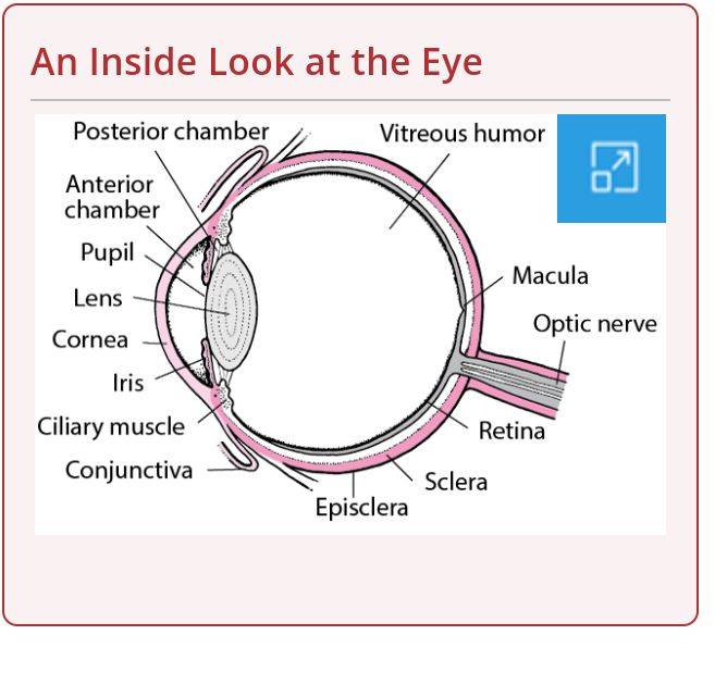
An Inside Look at the Eye
An Inside Look at the Eye
The outer covering of the eyeball consists of a relatively tough, white layer called the sclera (or white of the eye).
Near the front of the eye, in the area protected by the eyelids, the sclera is covered by a thin, transparent membrane (conjunctiva), which runs to the edge of the cornea. The conjunctiva also covers the moist back surface of the eyelids and eyeballs.
Light enters the eye through the cornea, the clear, curved layer in front of the iris and pupil. The cornea serves as a protective covering for the front of the eye and also helps focus light on the retina at the back of the eye.
After passing through the cornea, light travels through the pupil (the black dot in the middle of the eye).
The iris—the circular, colored area of the eye that surrounds the pupil—controls the amount of light that enters the eye. The iris allows more light into the eye (enlarging or dilating the pupil) when the environment is dark and allows less light into the eye (shrinking or constricting the pupil) when the environment is bright. Thus, the pupil dilates and constricts like the aperture of a camera lens as the amount of light in the immediate surroundings changes. The size of the pupil is controlled by the action of the pupillary sphincter muscle and dilator muscle.
Behind the iris sits the lens. By changing its shape, the lens focuses light onto the retina. Through the action of small muscles (called the ciliary muscles), the lens becomes thicker to focus on nearby objects and thinner to focus on distant objects.
The retina contains the cells that sense light (photoreceptors) and the blood vessels that nourish them. The most sensitive part of the retina is a small area called the macula, which has millions of tightly packed photoreceptors (the type called cones). The high density of cones in the macula makes the visual image detailed, just as a high-resolution digital camera has more megapixels.
Overview of the Eyes
Overview of the Eyes
VIDEO
Each photoreceptor is linked to a nerve fiber. The nerve fibers from the photoreceptors are bundled together to form the optic nerve. The optic disk, the first part of the optic nerve, is at the back of the eye.
The photoreceptors in the retina convert the image into electrical signals, which are carried to the brain by the optic nerve. There are two main types of photoreceptors: cones and rods.
Cones are responsible for sharp, detailed central vision and color vision and are clustered mainly in the macula.
Rods are responsible for night and peripheral (side) vision. Rods are more numerous than cones and much more sensitive to light, but they do not register color or contribute to detailed central vision as the cones do. Rods are grouped mainly in the peripheral areas of the retina.
The eyeball is divided into two sections, each of which is filled with fluid. The pressure generated by these fluids fills out the eyeball and helps maintain its shape.
The front section (anterior segment) extends from the inside of the cornea to the front surface of the lens. It is filled with a fluid called the aqueous humor, which nourishes the internal structures. The anterior segment is divided into two chambers. The front (anterior) chamber extends from the cornea to the iris. The back (posterior) chamber extends from the iris to the lens. Normally, the aqueous humor is produced in the posterior chamber, flows slowly through the pupil into the anterior chamber, and then drains out of the eyeball through outflow channels located where the iris meets the cornea.
The back section (posterior segment) extends from the back surface of the lens to the retina. It contains a jellylike fluid called the vitreous humor.
Tracing the Visual Pathways
Nerve signals travel from each eye along the corresponding optic nerve and other nerve fibers (called the visual pathway) to the back of the brain, where vision is sensed and interpreted. The two optic nerves meet at the optic chiasm, which is an area behind the eyes immediately in front of the pituitary gland and just below the front portion of the brain (cerebrum). There, the optic nerve from each eye divides, and half of the nerve fibers from each side cross to the other side and continue to the back of the brain. Thus, the right side of the brain receives information through both optic nerves for the left field of vision, and the left side of the brain receives information through both optic nerves for the right field of vision. The middle of these fields of vision overlaps. It is seen by both eyes (called binocular vision).
An object is seen from slightly different angles by each eye so the information the brain receives from each eye is different, although it overlaps. The brain integrates the information to produce a complete picture.
Tracing the Visual Pathways
CLICK HERE FOR THE PROFESSIONAL VERSION
Muscles, Nerves, and Blood Vessels of the Eyes
Others Also Read
Also Of Interest
MManual
MSD and the MSD Manuals
Merck and Co., Inc., Kenilworth, NJ, USA (known as MSD outside of the US and Canada) is a global healthcare leader working to help the world be well. From developing new therapies that treat and prevent disease to helping people in need, we are committed to improving health and well-being around the world. The Manual was first published in 1899 as a service to the community. The legacy of this great resource continues as the MSD Manual outside of the United States and Canada. Learn more about our commitment to Global Medical Knowledge.
follow us on Facebookfollow us on Twitter
ABOUT
DISCLAIMER
PERMISSIONS
PRIVACY
COOKIE PREFERENCES
TERMS OF USE
LICENSING
CONTACT US
GLOBAL MEDICAL KNOWLEDGE
VETERINARY EDITION
SOCIAL MEDIA
MOBILE APPS
This icon serves as a link to download the eSSENTIAL Accessibility assistive technology app for individuals with physical disabilities. It is featured as part of our commitment to diversity and inclusion.
View Privacy Shield Verification StatusView APEC CBPR Privacy Certification Status
This website is certified by Health On the Net Foundation. Click to verify.This site complies with the HONcode standard for trustworthy health information: verify here.
© 2022 Merck Sharp & Dohme Corp., a subsidiary of Merck & Co., Inc., Kenilworth, NJ, USA


No comments yet
Be the first to share your thoughts!