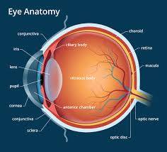
The structures and functions of the eyes are complex. The outer covering of the eyeball consists of a tough, white layer called the sclera, which is covered by a thin, transparent membrane called the conjunctiva. Light enters the eye through the cornea, which serves as a protective covering for the front of the eye and also helps focus light on the retina at the back of the eye. The iris controls the amount of light that enters the eye, allowing more light into the eye when the environment is dark and allowing less light when it is bright. The pupil is controlled by the action of the pupillary sphincter muscle and dilator muscle, and the lens focuses light onto the retina through the action of small muscles.
The retina is composed of cells that sense light (photoreceptors) and the blood vessels that nourish them. The most sensitive part of the retina is the macula, which has millions of tightly packed photoreceptors. Each photoreceptor is linked to a nerve fiber, which is bundled together to form the optic nerve. There are two main types of cones: cones and rods. The eyeball is divided into two sections, each of which is filled with fluid.
The front section (anterior segment) is filled with a fluid called the aqueous humor, which nourishes the internal structures. The anterior segment is divided into two chambers, the front (anterior) chamber extending from the cornea to the iris, and the back (posterior) chamber extends from the iris to the lens. The vitreous humor is produced in the posterior chamber, flows slowly through the pupil into the anterior chamber, and then drains out of the eyeball through outflow channels located where the iris meets the cornea. The optic nerve from each eye divides, and half of the nerve fibers from each side cross to the other side and continue to the back of the brain. The brain integrates the information to produce a complete picture.
Comments (0)
Be respectful and constructive in your comments.


No comments yet
Be the first to share your thoughts!